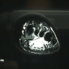
MRI showing an AVM on the plantar aspect of the foot
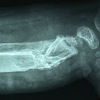
A 3-year-old child with deformity of ankle and distal tibia. AP and Lateral radiographs showing a destructive lesion in the bone with deformity. No surgical intervention was required. There was spontaneous resolution of the swelling and the bone remodeled completely.
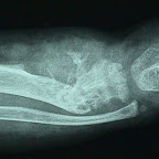
A 3-year-old child with deformity of ankle and distal tibia. AP and Lateral radiographs showing a destructive lesion in the bone with deformity. No surgical intervention was required. There was spontaneous resolution of the swelling and the bone remodeled completely.
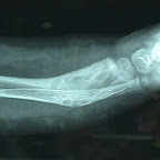
A 3-year-old child with deformity of ankle and distal tibia. AP and Lateral radiographs showing a destructive lesion in the bone with deformity. No surgical intervention was required. There was spontaneous resolution of the swelling and the bone remodeled completely.

A 3-year-old child with deformity of ankle and distal tibia. AP and Lateral radiographs showing a destructive lesion in the bone with deformity. No surgical intervention was required. There was spontaneous resolution of the swelling and the bone remodeled completely.

Soft tissue swelling on the lateral aspect of the thigh. MRI revealed a T2 enhancing soft tissue mass typical of AVM