
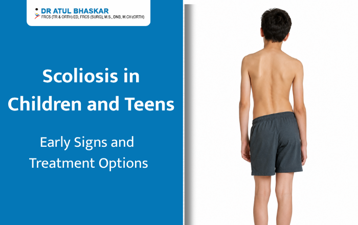
Scoliosis in children and teens is a condition in which the spine develops an abnormal sideways curve. It often becomes noticeable during the rapid growth that occurs around puberty. Some curves are mild and only need regular monitoring, while others may progress as a child grows and require treatment. Recognising the early signs can help parents seek an assessment at the right time. Treatment depends on factors such as the degree of spinal curvature, the child’s age, remaining growth, symptoms and whether the curve is changing over time. What Is Scoliosis in Children and Teens? Scoliosis is more than simply having poor posture. The spine may curve to one side and can also rotate, making the back appear uneven. The curve may resemble a “C” or “S” shape. The most common type in adolescents is adolescent idiopathic scoliosis, where an exact cause cannot be identified. It most commonly develops from around age 10 through the teenage years. Other forms can be associated with conditions affecting the muscles and nerves or abnormalities present in the spine from birth. Many children with mild scoliosis have no pain or obvious symptoms. This is why parents may notice changes in posture or body symmetry before the child complains of a problem. Early Signs of Scoliosis in Children and Teens Parents can look for changes in the child’s back, shoulders and waist, particularly during growth spurts. Common signs include: One shoulder appearing higher than the other One shoulder blade becoming more prominent An uneven waistline One hip appearing higher or more prominent The head appearing off-centre in relation to the hips Clothes hanging unevenly The child’s body appearing to lean to one side An abnormal curve becoming more visible when the child bends forward These signs do not necessarily mean that a child has scoliosis. Postural differences and other conditions can produce similar appearances. A healthcare professional can determine whether a spinal curve is actually present. Back pain is not always present in adolescent scoliosis. However, significant pain, particularly when accompanied by other symptoms, should be medically evaluated rather than automatically attributed to scoliosis. How Is Scoliosis Diagnosed? A doctor generally begins with a physical examination, looking at the child’s posture, shoulder and hip alignment and the appearance of the spine when standing and bending forward. If scoliosis is suspected, imaging may be recommended to evaluate the spine and measure the degree of curvature. The angle of the curve, together with the child’s skeletal maturity and growth potential, helps guide treatment decisions. Regular follow-up can be particularly important while a child is still growing because some curves can progress during periods of rapid growth. Treatment Options for Scoliosis in Children and Teens There is no single treatment that is appropriate for every child. Management is generally based on the severity and progression of the curve and how much growth remains. 1. Observation Mild curves may not require active treatment. Instead, the doctor may recommend regular examinations and, when appropriate, repeat X-rays to check whether the curve is changing. The Scoliosis Research Society generally describes observation as an option for curves below approximately 25 to 30 degrees, although the decision depends on the individual child. Observation does not mean ignoring scoliosis. It means monitoring the child’s spine carefully so treatment can be considered if the curve progresses. 2. Bracing A scoliosis brace may be recommended for a growing child with a moderate curve that has a meaningful risk of progression. Bracing is intended primarily to slow or prevent further worsening while the child continues to grow. The type of brace and recommended wearing schedule depend on the child’s curve and individual circumstances. For children who need bracing, following the prescribed wearing schedule can influence how effective the treatment is. 3. Surgery Surgery is considered for selected children with severe or significantly progressive curves, particularly when non-surgical treatment is not sufficient or the curve is unlikely to respond to bracing. For adolescent idiopathic scoliosis, spinal fusion with instrumentation is commonly used when surgery is indicated. The procedure aims to prevent further progression and correct the curve to an appropriate degree. Surgery is not required for most children with mild scoliosis. The decision should be made after considering the child’s growth, curve pattern, progression and overall health. Can Scoliosis Get Worse During Growth? Yes. A spinal curve can progress as a child grows, particularly during periods of rapid growth. This is one reason early assessment and follow-up are important. A child who has been diagnosed with scoliosis may therefore need periodic assessment until growth is substantially complete. The frequency of follow-up depends on the severity of the curve and the child’s stage of growth. When Should You Consult a Doctor? Parents should consider arranging an evaluation if they notice: Uneven shoulders or hips A visible spinal curve One shoulder blade becoming more prominent Persistent or significant back pain A noticeable change in posture during a growth spurt Increasing asymmetry of the back or waist A previously diagnosed spinal curve that appears to be changing Early evaluation does not automatically mean that a child will need a brace or surgery. In many cases, the first step is simply confirming whether scoliosis is present and monitoring its development. Frequently Asked Questions 1. Can scoliosis in children be corrected? Treatment can control progression and, depending on the severity and treatment used, improve the spinal curve. Mild curves may simply be monitored, while bracing or surgery may be considered for more significant or progressive curves. 2. Does scoliosis in teenagers always require a brace? No. Bracing is generally considered for selected growing children with curves at a level where progression is a concern. Mild curves may only require observation. 3. Is scoliosis caused by poor posture? Usually, no. Adolescent idiopathic scoliosis is not simply the result of sitting incorrectly or having poor posture. In the most common form, the exact cause is unknown. 4. Can children with scoliosis play sports? Many children with scoliosis can remain physically active. However, activity recommendations
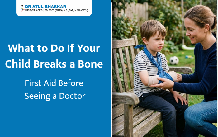
What to Do If Your Child Breaks a Bone: First Aid Before Seeing a Doctor
A snapped bone in your child is one of those moments that makes every parent’s stomach drop. One second they’re jumping off the monkey bars, and the next they’re crying in pain and refusing to move their arm. It’s scary, but here’s the good news: knowing what to do in the first few minutes can make a real difference in your child’s comfort and recovery. This guide walks you through exactly what to do — and what not to do — if you suspect your child has broken a bone. How to Tell If It’s Actually Broken Not every fall results in a fracture, and it’s not always obvious right away. Here are some signs that point to a possible broken bone rather than a simple bruise or sprain: Visible swelling or deformity (the limb looks bent or out of place) Severe pain that doesn’t ease up after a few minutes Inability to move or bear weight on the injured area A popping or snapping sound at the time of injury Numbness or tingling near the injury site If you notice any of these signs, it’s safer to assume it’s a fracture until a doctor confirms otherwise. Step 1: Stay Calm and Keep Your Child Calm Children pick up on panic instantly. Take a breath before you react. Speak in a steady, reassuring voice. Your calmness will help your child stay still, which matters because unnecessary movement can worsen the injury. Step 2: Don’t Move the Injured Area This is one of the most important rules of fracture first aid. Avoid trying to straighten a bent limb or forcing your child to move the injured part. Let it stay exactly as it is until it’s properly supported. Step 3: Immobilize the Area Immobilization prevents further damage and reduces pain. Here’s how to do it safely at home: Use a rolled-up towel, magazine, or a rigid object as a makeshift splint Gently place it alongside the injured limb without forcing it into position Secure it loosely with a bandage, scarf, or cloth strips — not too tight Support the area above and below the suspected break If it’s an arm injury, a simple sling made from a piece of cloth can help keep it stable while you head to the hospital. Step 4: Apply a Cold Compress Wrap ice or a cold pack in a thin cloth and place it gently near (not directly on) the injured area for about 15-20 minutes. This helps reduce swelling and numbs the pain slightly. Never apply ice directly to the skin, as it can cause frostbite, especially in young children. Step 5: Elevate the Injury If Possible If the break is in an arm or leg, try to keep it elevated slightly above heart level, without moving the injured limb itself. This helps control swelling. If your child is in too much pain to allow this, don’t force it. Step 6: Manage Pain Carefully You can give an age-appropriate dose of paracetamol (acetaminophen) if your child is in pain, following the dosage instructions for their age and weight. Avoid giving ibuprofen or aspirin unless a doctor has already advised it, especially if there’s any bleeding or if surgery might be needed soon. Step 7: Watch for Warning Signs While heading to the doctor, keep an eye out for: The skin turning pale, blue, or unusually cold Loss of sensation in fingers or toes The bone piercing through the skin (open fracture) Excessive bleeding If any of these occur, treat it as a medical emergency and get to an emergency room immediately rather than waiting for a regular appointment. What NOT to Do Don’t try to “pop” the bone back into place Don’t give your child food or water if surgery might be needed Don’t ignore the injury just because your child stops crying — pain tolerance varies, and shock can mask symptoms Don’t massage or apply pressure directly on the injury When to Head Straight to the Hospital Some fractures need urgent attention. Go to the emergency room right away if: The bone is visibly out of place or piercing the skin Your child has lost feeling in the limb There’s heavy bleeding The pain is severe and unmanageable You notice signs of shock, like pale skin, rapid breathing, or dizziness For less severe injuries, a visit to a pediatric orthopedic specialist within a few hours is usually appropriate. Helping Your Child Feel Safe Beyond the physical first aid, don’t underestimate the emotional side of this experience. A calm tone, a comforting hand, and simple explanations (“The doctor is going to help make it feel better”) go a long way in helping your child stay cooperative and less frightened. FAQs: Quick Answers to Common Questions Q1: Can I tell if a bone is broken just by looking at it? Not always. Some fractures, especially in young children, don’t show obvious deformity. Persistent pain, swelling, or refusal to use the limb is enough reason to get it checked. Q2: Should I try to splint the injury myself before going to the hospital? Yes, a simple, gentle splint can help stabilize the area during transport, as long as you don’t force the limb into a new position. Q3: How soon should my child see a doctor after a suspected fracture? As soon as possible — ideally within a few hours. Delayed treatment can affect healing, especially in growing bones. Q4: Is it okay to give my child pain medicine before seeing a doctor? A dose of paracetamol appropriate for their age and weight is generally safe. Avoid other medications unless advised by a healthcare professional. Q5: Do children’s bones heal faster than adults’? Generally, yes. Children’s bones are still growing and tend to heal more quickly, but proper diagnosis and treatment are still essential to avoid complications. Have questions? We’re just a message away! Contact us on WhatsApp — Click Here to Chat with Us Conclusion First aid can help manage the situation, but it’s
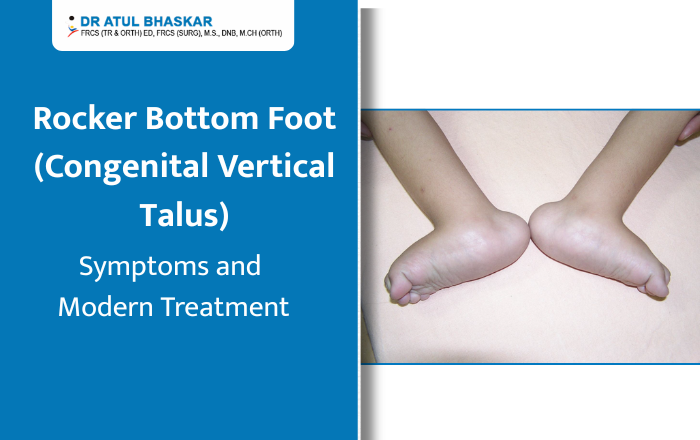
Rocker Bottom Foot (Congenital Vertical Talus): Symptoms and Modern Treatment
Imagine holding your newborn for the very first time and noticing that one or both of their tiny feet looks unusual — the sole curves outward like the bottom of a rocking chair, and no matter how gently you try to move it, it stays stiff. Your heart sinks. You have questions, and you need answers. If this sounds familiar, you may be looking at a condition called rocker bottom foot, medically known as Congenital Vertical Talus (CVT). It sounds intimidating, but here is the truth — this condition is rare, it is diagnosable, and most importantly, it is very treatable, especially when caught early. This article will walk you through everything you need to know, in plain, simple language. What Is Rocker Bottom Foot? Rocker bottom foot is a congenital (present at birth) deformity where the talus bone in the foot points straight downward instead of sitting at its natural angle. This causes the sole of the foot to bow outward, creating that distinctive rounded, rocker-like shape. Unlike a simple flat foot — which is flexible and often harmless — congenital vertical talus is rigid. The foot cannot be moved into a normal position by hand. It affects roughly 1 in 10,000 newborns and occurs in both feet in about half of all cases. What Causes It? In many children, rocker bottom foot occurs on its own without any clear reason. However, it is sometimes linked to: Neuromuscular conditions like spina bifida or arthrogryposis Chromosomal abnormalities such as Trisomy 13 or Trisomy 18 Connective tissue disorders In some cases, it can even be spotted during a prenatal ultrasound. If your doctor notices something unusual before birth, early referral to a pediatric orthopedic specialist is strongly recommended. Symptoms: What Should You Look For? Spotting congenital vertical talus early makes a significant difference in how smoothly treatment goes. Here are the key signs: The sole of the foot is visibly rounded or convex — like the underside of a rocking chair The foot feels stiff and rigid, even when you try to gently flex it The heel points downward while the front of the foot turns upward and outward There may be a bony bump visible on the inner side of the foot Standard baby shoes or booties simply do not fit properly As the child grows older and begins to walk, untreated rocker bottom foot can cause limping, pain, and the development of thick calluses on the curved sole of the foot. How Is It Diagnosed? A pediatric orthopedic surgeon will first perform a careful physical examination, checking how the foot moves and feels. They will then use X-rays — including stress-position views — to confirm the vertical position of the talus bone and rule out other conditions like flexible flatfoot or oblique talus. The earlier the diagnosis, the better. Babies diagnosed before 6 months of age respond far better to non-surgical treatment because their joints and soft tissues are still highly flexible. Modern Treatment Options for Rocker Bottom Foot The Dobbs Casting Method The biggest shift in congenital vertical talus treatment over the past two decades has been the Dobbs casting technique. Before this method, surgery was almost always the first option. Today, it is the gold standard first-line approach worldwide. Here is how it works: The surgeon gently manipulates the foot into a better position during each clinic visit, then applies a series of plaster casts — typically over 4 to 6 weeks — to gradually move the bones toward their correct alignment. Studies show this approach achieves meaningful correction in up to 70–80% of cases. After casting, a small procedure under anesthesia — a percutaneous pin fixation — is usually performed to hold the talus in place. This is far less involved than traditional open surgery and carries a much shorter recovery time. When Surgery Is Needed For older children, more severe cases, or situations where casting alone has not achieved full correction, surgical correction may be recommended. This typically involves tendon lengthening and stabilization of the joint, and is most effective when performed between 6 and 18 months of age. After Treatment: Bracing and Follow-Up Treatment does not stop after casting or surgery. Children are placed in a foot abduction brace — similar to what is used after clubfoot treatment — to maintain the corrected position during growth. Physiotherapy exercises also help strengthen the foot muscles over time. Regular follow-ups every six months through the growing years are essential to catch any signs of recurrence early. What Happens If It Goes Untreated? This is important for every parent to understand. Rocker bottom foot does not resolve on its own. Without treatment, children grow up with a painful, rigid deformity that makes walking difficult, limits physical activity, and may eventually require far more complex surgery in adulthood. Early action is not optional — it is everything. Trust Your Child’s Feet to the Right Hands If you suspect your child has rocker bottom foot or congenital vertical talus, do not wait. In Mumbai, Dr. Atul Bhaskar, a Pediatric Orthopedic Surgeon with over 33+ years of experience, has helped countless families navigate this condition with skill and compassion. From diagnosis through recovery, Dr. Bhaskar’s approach puts your child’s long-term mobility and quality of life first. Name – Dr. Atul R Bhaskar . PAEDIATRIC ORTHOPAEDIC SURGEON Address: B Wing, Laxmi Business Park, Room No. 114, Sab T V – Kalinga Ln, off New Link Road, Laxmi Industrial Estate, Suresh Nagar, Andheri West, Mumbai, Maharashtra 400053 Phone: 98216 22992 Book a consultation with Dr. Atul Bhaskar today — because every child deserves to walk, run, and play without limits.
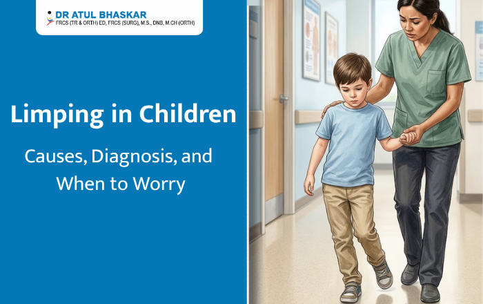
Limping in Children: Causes, Diagnosis, and When to Worry
If you’ve noticed your child limping lately, you’re probably worried. And that’s completely normal. As a parent, watching your child walk differently — dragging one leg, favouring one side, or wincing with each step — is unsettling. But here’s the reassuring truth: limping in children is more common than most parents realise, and many causes are simple and treatable. That said, some causes do need urgent attention. This blog will walk you through everything you need to know — from common reasons to serious warning signs — so you can make the right call at the right time. Why Is My Child Limping? Understanding the Basics A limp in a child simply means their walking pattern has changed. It can happen because of pain, weakness, or a structural problem in the leg, hip, knee, or foot. Doctors broadly classify limps into three types — pain-related (antalgic), weakness-related (Trendelenburg), or mechanical (like a leg length difference).Age plays a big role too. A limping toddler is often looked at differently than a limping teenager, because different conditions tend to show up at different stages of development. 9 Most Common Causes of Limping in Children 1. Transient Synovitis — The Most Frequent CulpritThis is the single most common reason for limping in children aged 3–10. It’s a temporary inflammation of the hip joint, often following a cold or viral infection. The good news? It usually gets better on its own within 1–4 weeks with rest.2. Injury or TraumaSprains, minor fractures, or muscle pulls are obvious causes. One commonly missed injury is “toddler’s fracture” — a small crack in the shin bone that can happen even without a noticeable fall.3. Legg-Calve-Perthes (LCP) DiseaseIn this condition, the blood supply to the ball of the hip joint is temporarily disrupted, causing the bone to weaken. It affects about 1 in 1,200 children, mostly boys between 4–10 years. A child limping without pain is often the first — and only — sign.4. Septic Arthritis — A Medical EmergencyThis is a bacterial infection inside a joint. If your child is limping with a fever, refuses to move their leg, and is in severe pain — go to the emergency room immediately. Septic arthritis can permanently damage a joint within hours if not treated.5. Slipped Capital Femoral Epiphysis (SCFE)This condition affects teenagers, particularly overweight boys. The “ball” of the hip slips off its normal position. Many children with SCFE complain of knee pain — but the real problem is in the hip. Surgery is almost always needed.6. Growing Pains vs. Juvenile ArthritisTrue growing pains cause nighttime aching in both legs and do not cause a limp. If your child is limping, has joint swelling, or feels stiff in the mornings for more than 6 weeks, juvenile arthritis should be evaluated.7. Flat Feet or Hip DysplasiaThese structural conditions often cause a painless limp in young children and are frequently missed. Early detection makes treatment far simpler.8. Osteomyelitis (Bone Infection)Similar to septic arthritis, this is a bone infection that causes fever, localised bone pain, and reluctance to walk. It needs prompt treatment with antibiotics or surgery.9. Rare but Serious: Bone Tumors and LeukemiaA persistent child limping — especially with night pain, fatigue, or unexplained weight loss — should never be ignored. Bone pain is a presenting symptom in roughly 25% of childhood leukemia cases. This is rare, but worth ruling out. 7 Red Flag Warning Signs You Should Never Ignore Do not wait. Contact your doctor immediately if your limping child shows any of these signs: Fever along with a limp — possible joint or bone infection Refusal to bear any weight on the leg Swollen, red, or warm joint Limp lasting more than 1–2 weeks Night pain that wakes the child from sleep Unexplained tiredness or weight loss Any limp in an infant or newborn — always abnormal How Do Doctors Diagnose the Cause of a Limp? When you bring your limping child to a doctor, here’s what typically happens: Physical examination comes first. The doctor watches your child walk, checks the range of motion in the hip and knee, looks for swelling, and compares leg lengths. Investigations are chosen based on what’s suspected. X-rays pick up fractures, LCP disease, and SCFE. An ultrasound can detect fluid in the hip joint — a key difference between harmless transient synovitis and dangerous septic arthritis. Blood tests (CRP, ESR, white blood cell count) check for infection or inflammation. An MRI is used when soft tissue or early avascular necrosis needs to be assessed. Doctors also use the Kocher Criteria — four clinical markers — to quickly identify children at high risk for septic arthritis. The more criteria a child meets, the greater the urgency. Treatment: From Rest to Surgery Treatment depends entirely on the cause. Rest and medication work well for transient synovitis and minor injuries — most children recover fully at home. Physiotherapy and bracing help with flat feet, mild hip dysplasia, and certain stages of LCP disease. Surgery becomes necessary for SCFE, severe LCP disease, septic arthritis (joint drainage), and untreated hip dysplasia. The earlier surgery is done when needed, the better the outcome for the growing joint. Special Scenarios Parents Often Search For My child limps only in the morning — Morning stiffness lasting more than 30 minutes alongside a limp in a child points toward juvenile idiopathic arthritis. My child limps after sports — This often suggests stress fractures or Osgood-Schlatter disease in active teenagers. My child limps but doesn’t complain of pain — A painless limp is a structural clue, not a reassuring one. Conditions like LCP disease and hip dysplasia are often completely painless in the early stages. My toddler just started limping — Could be a toddler’s fracture, hip dysplasia, or early transient synovitis. Always worth a check-up. FAQs: Limping in Children Should I go to the ER if my child is limping?Yes — if there is fever, refusal to walk, or a swollen joint. For a mild limp with no other symptoms, a doctor visit within
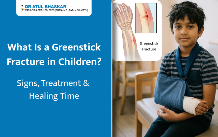
What Is a Greenstick Fracture in Children? Signs, Treatment & Healing Time
Your child was playing outside, tripped, and landed awkwardly — now they’re crying and holding their arm. You rush over, expecting the worst, but there’s no obvious break. Could it still be a fracture? Yes, it absolutely could. It might be a greenstick fracture — one of the most common yet misunderstood bone injuries in children. As a parent, understanding what a greenstick fracture in children is, how to spot it early, and what treatment looks like can save your child from unnecessary pain and complications. Let’s break it all down in simple, clear terms. What Is a Greenstick Fracture? A greenstick fracture is an incomplete bone break where the bone bends and cracks on one side but doesn’t break all the way through. Picture trying to snap a fresh green twig — it bends and splits rather than breaking cleanly. That’s exactly what happens inside your child’s bone. This type of fracture happens almost exclusively in children because kids’ bones are softer and more flexible than adult bones. The younger the child, the more pliable their bones — which is why greenstick fractures are most common in children under 10 years of age. Where Do Greenstick Fractures Usually Occur? The most common locations include: Forearm (most frequent — from falling on an outstretched hand) Collarbone (clavicle) Shinbone (tibia) Upper arm (humerus) Signs and Symptoms — How Do You Know? Because the bone doesn’t fully snap, a greenstick fracture can easily be mistaken for a sprain. Here’s what to look for: Pain at the injury site, especially when touched Swelling around the affected area Mild bowing or bending of the limb — it may look slightly off Bruising appearing within 24–48 hours Child refusing to use the arm or leg — a classic warning sign in young kids Tenderness that doesn’t improve after a few hours When Should You See a Doctor Immediately? Take your child to a doctor right away if: The limb looks visibly bent or deformed There is severe, continuous pain Fingers or toes below the injury feel numb or look pale or bluish Your child completely refuses to walk (for leg injuries) Never assume it’s just a sprain. In growing children, untreated fractures can affect long-term bone development. How Is a Greenstick Fracture Diagnosed? A doctor will first do a physical examination — checking for swelling, tenderness, and movement. An X-ray is then used to confirm the greenstick fracture diagnosis. In some cases, two different angled X-ray views are needed because the fracture line can be very subtle. Rarely, an MRI may be ordered if the fracture is near a growth plate. Greenstick Fracture Treatment — What to Expect The good news is that most greenstick fractures heal without surgery. Treatment depends on how much the bone has bent. 1. Casting A plaster or fibreglass cast is the most common treatment. It holds the bone still while it heals and prevents the bend from getting worse. Most children wear a cast for 4 to 8 weeks. 2. Splinting For mild cases, a splint may be used initially — especially while swelling is still present — before a proper cast is applied. 3. Realignment (Closed Reduction) If the bone is bent more than 15–20 degrees, the doctor will gently straighten it before casting. This is done under sedation so your child feels no pain during the process. 4. Surgery Surgery is rarely needed for greenstick fractures. It is only considered in very severe cases where the bone cannot be properly aligned otherwise. At-Home Care Tips Elevate the injured limb for the first 48 hours Use an ice pack (wrapped in cloth) for 15–20 minutes at a time Give pain relief as directed by your doctor Keep the cast completely dry Greenstick Fracture Healing Time Children’s bones heal much faster than adults’ because they are still actively growing. Here’s a simple guide: Child’s Age Healing Time Under 2 years 3–4 weeks 2–5 years 4–6 weeks 6–10 years 6–8 weeks 10+ years 8–12 weeks After the cast comes off, mild stiffness and weakness are completely normal. Most children return to sports and full activity within 2–4 weeks after cast removal. FAQ Q1. Can a child walk with a greenstick fracture? If the fracture is in the leg, some walking may be possible — but it can make the injury worse. Always get an X-ray before letting your child put weight on an injured leg. Q2. Does a greenstick fracture need a cast? Almost always, yes. Without a cast, the bone can bow further or heal in a bent position, causing long-term issues. Q3. Will it affect my child’s bone growth? When diagnosed and treated on time, it typically does not affect growth. However, if the growth plate is involved, your doctor will monitor the healing closely. Trust Your Child’s Recovery to the Right Hands A greenstick fracture may sound less serious than a full break — but it is still a real fracture that needs proper treatment. Catching it early and treating it correctly makes all the difference. Dr. Atul Bhaskar — Paediatric Orthopaedic Surgeon, Mumbai When it comes to your child’s bones, you want a doctor who has seen it all — and treated it all. Dr. Atul Bhaskar is one of Mumbai’s most trusted Paediatric Orthopaedic Surgeons with 33+ years of experience treating children’s bone injuries, including greenstick fractures. Families across Mumbai rely on Dr. Bhaskar for his calm, child-friendly approach and his deep knowledge of growing bones. ✅ 33+ years in paediatric orthopaedics ✅ Thousands of successful fracture treatments in children ✅ Gentle, child-friendly approach ✅ Trusted by Mumbai families for over three decades Your child’s bones are still growing — make sure they heal right. Book a consultation with Dr. Atul Bhaskar today and give your child the care their recovery truly deserves.
Delayed Walking in Toddlers: Causes, Signs & Treatment
You dressed them up, you cheered them on, and you waited. But while other toddlers at the park are wobbling around on two feet, your little one is still crawling — and that quiet worry in the back of your mind is getting louder. First, take a breath. Delayed walking in toddlers is more common than most parents realize, and in many cases, it is completely treatable. This article will walk you through everything you need to know — from causes and warning signs to treatment options — so you can make the best decision for your child. What Age Should Toddlers Start Walking? Most babies take their first steps somewhere between 9 and 12 months and are walking confidently by 14 to 15 months. The medical community considers 18 months the upper limit of the normal range. If your child is not walking by 18 months, that is the point where a specialist visit becomes important — not a reason to panic, but definitely a reason to act. 7 Common Causes of Delayed Walking in Toddlers Understanding why delayed walking in toddlers happens is the first step toward finding the right solution. 1. Low Muscle Tone (Hypotonia) Some children are born with weaker muscle tone, making it harder to bear weight on their legs. You may notice floppy limbs or difficulty sitting upright for long periods. 2. Hip Dysplasia This is a condition where the hip joint does not form correctly, making it painful or difficult for a toddler to stand and walk. It is one of the most commonly missed causes of walking delay in babies and needs early orthopedic attention. 3. Club Foot or Flat Feet Structural problems in the foot — such as club foot or severely flat feet — can affect balance and make walking uncomfortable. Most of these conditions respond very well to early treatment. 4. Neurological Conditions Conditions like cerebral palsy or spina bifida can affect the brain’s ability to send signals to the muscles. Toddlers with delayed walking due to neurological causes benefit greatly from early diagnosis. 5. Vitamin D Deficiency & Rickets This is surprisingly common in India, especially in cities where children spend less time outdoors. Soft or weak bones caused by Vitamin D deficiency can directly delay a child’s ability to walk. 6. Developmental Coordination Disorder Some children struggle with coordination between the brain and body. Their muscles are fine, but the communication between the two needs support through therapy. 7. Environmental Factors Too much time in a pram or baby walker, and not enough floor time, can slow down natural motor development. The American Academy of Pediatrics (AAP) strongly advises against baby walkers as they can actually delay independent walking. Warning Signs You Should Not Ignore Not every late walker has a medical problem — but certain signs deserve prompt attention: Your toddler is not walking by 18 months They walk only on their toes after 24 months One leg appears stronger or more active than the other Their legs seem unusually stiff or unusually floppy They had started showing signs of walking but then stopped progressing If you notice any of the above, do not wait to “see how it goes.” Early evaluation makes a real difference in outcomes. How Is Walking Delay Diagnosed? A pediatric orthopedic surgeon will typically begin with a physical examination — checking muscle tone, joint flexibility, leg symmetry, and reflexes. Depending on the findings, they may recommend: X-rays to check for hip dysplasia or bone abnormalities Blood tests to check Vitamin D and calcium levels MRI scans if a neurological cause is suspected The goal is to identify the root cause so that treatment targets the right problem. Treatment Options for Delayed Walking in Toddlers Most causes of delayed walking in toddlers are treatable — especially when caught early. Physiotherapy This is often the first line of treatment. A trained physiotherapist uses play-based exercises to strengthen your child’s legs, hips, and core. Most children respond well and begin making visible progress within weeks. Orthotic Devices For children with flat feet or balance issues, supportive footwear or ankle-foot orthotics (AFOs) can provide the stability they need to walk with confidence. Nutritional Treatment If Vitamin D deficiency is the cause, targeted supplementation often leads to quick and noticeable improvement in strength and movement. Surgery — Only When Truly Needed In cases like hip dysplasia or club foot that have not responded to other treatments, surgical correction may be recommended. Modern techniques are safe, and recovery with proper physiotherapy is usually smooth. Take the First Step — Consult Dr. Atul Bhaskar If your child is showing signs of delayed walking, do not wait and wonder. Every month matters when it comes to a growing child’s development. Dr. Atul Bhaskar, Pediatric Orthopedic Surgeon based in Andheri West, Mumbai, brings over 33+ years of experience in diagnosing and treating walking delays, hip dysplasia, club foot, and other pediatric bone and joint conditions. Families across Mumbai trust Dr. Bhaskar for his clear, honest guidance and genuine care for every child he sees. 📍 Andheri West, Mumbai 👨⚕️ 33+ Years | Pediatric Orthopedic Surgery 👉 Book a consultation with Dr. Atul Bhaskar today — because your child deserves to run, jump, and play without limits.
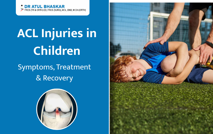
ACL Injuries in Children: Symptoms, Treatment & Recovery
If your child suddenly grabs their knee during a soccer game or basketball practice, your heart might skip a beat. Knee injuries are scary, especially when they happen to our little ones. One of the most common serious knee injuries in young athletes is an ACL injury in children. The ACL, or Anterior Cruciate Ligament, is a strong band of tissue that keeps the knee stable. When it tears, it can sideline your child from sports and cause real pain. The good news? With the right care and treatment, most kids recover well and get back to doing what they love. In this guide, we’ll walk you through everything you need to know about pediatric ACL tears—from spotting the signs to understanding treatment options and helping your child heal. What Is an ACL Injury? The ACL is one of four main ligaments in the knee. It connects the thigh bone to the shin bone and stops the shin from sliding too far forward. It also helps keep your knee stable when you twist or turn. ACL injuries in children are becoming more common than they used to be. More kids are playing competitive sports year-round, which means more chances for injury. Sports like soccer, basketball, football, and gymnastics carry the highest risk. Children’s knees are different from adult knees because they’re still growing. They have growth plates—areas of developing bone tissue near the ends of long bones. This means doctors must be extra careful when treating pediatric ACL injuries to protect normal growth. How Do ACL Injuries Happen in Kids? ACL tears in children usually happen during sports or active play. Common causes include: Suddenly stopping or changing direction while running Landing awkwardly from a jump Getting hit directly on the knee Pivoting with the foot planted firmly on the ground Girls are at higher risk than boys for ACL injuries. This might be due to differences in muscle strength, knee alignment, and how they land from jumps. Kids who specialize in just one sport at a young age or who play on multiple teams may also face higher injury risks due to overuse and fatigue. Recognizing ACL Injury Symptoms in Your Child How do you know if your child has an ACL tear? Watch for these warning signs: Right after the injury: A popping sound or sensation in the knee Sudden, severe pain Rapid swelling (usually within 2-12 hours) Trouble putting weight on the leg Feeling like the knee is unstable or “giving out” In the days following: Ongoing knee pain Difficulty walking, especially on stairs Limited knee movement Continued swelling Not wanting to run or play If your child shows these symptoms after a knee injury, schedule an appointment with a doctor soon. Early diagnosis helps protect the knee from further damage. How Doctors Diagnose ACL Injuries When you visit the doctor, they’ll examine your child’s knee, checking for swelling, tenderness, and stability. They may perform special tests by gently moving the knee in different directions. An MRI scan is usually needed to confirm an ACL tear in children. This imaging test shows a clear picture of the ligament and reveals if there’s any other damage to the knee. X-rays might also be taken to check for broken bones and to see how much your child’s bones have grown. Treatment Options for Pediatric ACL Injuries Treatment depends on several factors: your child’s age, how active they are, and how severe the injury is. Non-Surgical Treatment Some children, especially younger ones or those with partial tears, may not need surgery right away. Non-surgical treatment includes: Rest and activity modification Physical therapy to strengthen the muscles around the knee Wearing a knee brace for support Avoiding sports that involve cutting, pivoting, or jumping The challenge is that a torn ACL doesn’t heal on its own. Without surgery, the knee may remain unstable, which can lead to more damage over time. Surgical Treatment For active kids who want to return to sports, ACL surgery is often recommended. The concern with younger children is protecting their growth plates during surgery. Today, surgeons use special techniques designed for growing kids. These methods reconstruct the ACL while avoiding or minimizing impact on growth plates. The surgery typically uses a graft (tissue taken from another part of the body or from a donor) to rebuild the torn ligament. Surgery for ACL injuries in children is usually done as an outpatient procedure, meaning your child can go home the same day. Recovery Timeline: What to Expect ACL recovery in children takes time and patience. Here’s a general timeline: Weeks 1-6: Focus on reducing swelling and pain. Your child will work on gentle knee movements and may use crutches. Weight-bearing increases gradually. Months 2-4: Physical therapy intensifies. Your child will do exercises to strengthen the leg muscles, improve balance, and increase flexibility. Months 4-9: Sport-specific training begins. Running, jumping, and agility drills are introduced carefully under supervision. Months 9-12: Most children can return to competitive sports around 9-12 months after surgery, once they pass specific strength and function tests. Every child heals at their own pace. Rushing back too soon increases the risk of re-injuring the knee. The Role of Physical Therapy Physical therapy is the backbone of ACL recovery. A good PT program helps your child regain strength, flexibility, and confidence. The therapist will guide your child through exercises that gradually rebuild the knee’s function. As a parent, your encouragement matters. Recovery is long, and kids can get frustrated. Celebrate small victories and remind them that taking time to heal properly means they can play safely for years to come. Can You Prevent ACL Injuries? While you can’t prevent every injury, you can reduce the risk: Enroll your child in training programs that teach proper jumping and landing techniques Make sure they do strength exercises for the legs, hips, and core Encourage rest days and playing multiple sports instead of specializing too early Ensure coaches prioritize safety and proper form Finding the Right Care for
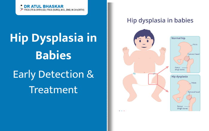
Hip Dysplasia in Babies: Early Identification & Treatment Guide for Parents
When you’re changing your baby’s diaper and notice one leg looks shorter than the other, or the skin folds don’t match up, it’s natural to feel worried. These could be early signs of hip dysplasia in babies—a condition where the hip joint doesn’t form properly. But here’s the good news: when spotted early, hip dysplasia is highly treatable. Most babies who receive timely care grow up to run, jump, and play just like other children. This guide will help you understand what to look for and when to seek help. What is Hip Dysplasia in Babies? Hip dysplasia in babies, also called Developmental Dysplasia of the Hip (DDH), happens when the hip joint doesn’t develop correctly. Think of a healthy hip as a ball sitting snugly in a socket. With hip dysplasia, the socket is too shallow, or the ball doesn’t fit properly. This can range from mild looseness to complete dislocation. About 1-2 babies per 1,000 are born with this condition. Girls are four times more likely to have it than boys. The condition can develop before birth, during delivery, or in the first year of life. 7 Warning Signs Every Parent Should Know Catching hip dysplasia early makes treatment easier. Watch for these signs during diaper changes: 1. Uneven skin folds – One thigh or buttock has more creases than the other 2. One leg looks shorter – When you bend both knees, one sits lower than the other 3. Limited hip movement – Difficulty spreading one leg to the side during diaper changes 4. Clicking sounds – You hear or feel a “clunk” when moving your baby’s legs 5. One foot turns out differently – The affected leg rotates outward more than the other 6. Uneven crawling or walking – Your child limps or favors one leg 7. Baby prefers one side – Doesn’t kick both legs equally or only rolls one direction Remember: Not all babies with hip dysplasia show obvious signs. This is why regular check-ups with your pediatrician matter. What Causes Hip Dysplasia? Several factors increase the risk of baby hip dysplasia: Breech position: Babies born bottom-first have higher risk Family history: Having a parent or sibling with hip dysplasia increases chances First-born babies: Less womb space can affect hip development Being female: Hormones make girls more susceptible Tight swaddling: Wrapping legs too straight can contribute to the problem You can help reduce risk by swaddling your baby with hips bent and free to move, and using baby carriers that support healthy hip positioning. How Doctors Check for Hip Dysplasia Your baby’s doctor will screen for hip dysplasia during regular visits. They’ll: Gently move your baby’s hips to check stability Look at leg length and skin folds Watch how your baby moves If something seems off, your doctor may recommend: Ultrasound (for babies under 6 months) – A safe, painless way to see the hip structure X-rays (for babies over 4-6 months) – Used once bones are more developed Studies show that early screening catches 90% of cases, leading to better results with simpler treatment. Treatment Options That Work The earlier hip dysplasia is found, the simpler the treatment. Here’s what to expect: ▸Pavlik Harness (For Babies Under 6 Months) This is the most common treatment. The soft harness holds your baby’s hips in the correct position for 6-12 weeks. Success rate? An impressive 90-95% when started early. Your baby can still move their legs and be held. Most parents say their little ones adjust within days. ▸Abduction Brace (For Older Babies) If your baby is over 6 months old, a stiffer brace might be needed. It keeps the legs in a “frog-leg” position and is worn for several months. ▸Closed Reduction If the harness doesn’t work, doctors can reposition the hip under anesthesia. Your baby will then wear a cast for 2-4 months. ▸Surgery (For Severe Cases) Reserved for complex situations or late diagnosis. With proper surgical treatment, children still have excellent long-term outcomes. ▸Will My Baby Walk Normally? Yes! With early treatment, over 95% of children with hip dysplasia lead completely normal, active lives. They participate fully in sports, dancing, and all childhood activities. Most children who wear a harness as infants show no lasting effects. You’d never know they had the condition. ▸Living with a Hip Harness Many parents worry about daily care, but it’s manageable: Diaper changes: Definitely possible—you’ll get the hang of it quickly Bathing: Your doctor will show you how to clean under the harness Sleep: Babies adapt fast and sleep comfortably Bonding: You can still cuddle, breastfeed, and comfort your baby normally When to See a Doctor Don’t wait if you notice: Any warning signs mentioned above Limping when your child starts walking Hip or leg pain in older children Any concerns about your baby’s hip development Hip dysplasia becomes harder to treat the longer it goes undiagnosed. Trust your instincts—if something feels wrong, get it checked. Get the Right Care in Mumbai If you’ve noticed signs of hip dysplasia in your baby, or your child has been diagnosed, getting proper medical care is crucial. Dr. Atul Bhaskar, a Paediatric Orthopaedic Surgeon in Mumbai, specializes in treating hip dysplasia and other childhood conditions. With extensive experience in both harness treatment and surgical options when needed, Dr. Bhaskar helps families navigate this journey with confidence. Why families choose Dr. Atul Bhaskar: Specialized training in children’s hip conditions Experience with Pavlik harness fitting and monitoring Surgical expertise for complex cases Clear explanations that parents understand Supportive care throughout your child’s growth Your baby’s hip health is important. Early diagnosis and proper treatment mean your little one can grow up healthy, active, and pain-free. Schedule a consultation with Dr. Atul Bhaskar today to get the answers and treatment your family needs.
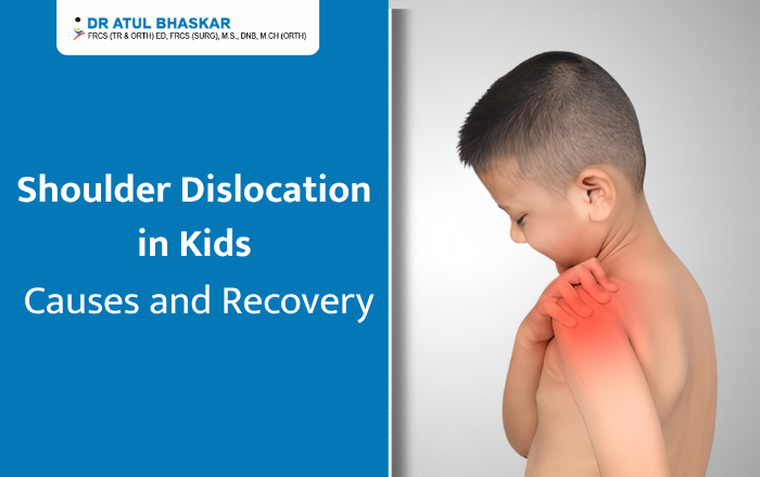
Shoulder Dislocation in Kids: Causes, Symptoms, and Recovery Timeline
Shoulder Dislocation in Kids: Causes, Symptoms, and Recovery Timeline When your child suddenly grabs their shoulder in pain during a game or after a fall, your heart skips a beat. Is it serious? What should you do? If you’re searching for answers about shoulder dislocation in kids, you’re in the right place. Shoulder dislocations can happen to children, especially active ones who love sports and outdoor play. While it’s more common in teenagers than younger kids, knowing what to look for and how to respond can make all the difference. Let’s break down everything you need to know in simple terms. What Is a Shoulder Dislocation? Think of your child’s shoulder as a ball sitting in a shallow cup. The upper arm bone (the ball) normally sits snugly in the shoulder socket (the cup). A dislocation happens when that ball pops completely out of the socket. This is different from a shoulder separation, which affects a different part of the shoulder where the collarbone meets the shoulder blade. It’s also more serious than a strain or sprain. Children’s shoulders are still growing, which actually makes complete dislocations less common in younger kids compared to teens. Their bones and joints are more flexible, but when a dislocation does happen, it needs immediate medical attention. What Causes Shoulder Dislocation in Children? Sports InjuriesMost shoulder dislocations in kids happen during sports, especially: Contact sports like football, hockey, or rugby Activities with potential falls like gymnastics or wrestling Overhead sports such as swimming, baseball, or volleyball A direct hit, awkward landing, or extreme arm movement can force the shoulder out of place. Falls and Accidents Outside of sports, common causes include: Playground accidents and falls Bicycle or skateboard crashes Landing on an outstretched arm during play Car accidents (less common but possible) Joint LoosenessSome children have naturally looser joints, making dislocations more likely. This can run in families or be part of certain medical conditions affecting connective tissue. How to Tell If Your Child’s Shoulder Is Dislocated Look for These Signs: ▹Pain and Position Sudden, intense pain in the shoulder Your child can’t move their arm They hold their arm in an odd position away from their body They may tell you something “popped out” ▹Visible Changes The shoulder looks different or deformed One shoulder appears lower than the other Swelling develops quickly Bruising may appear Important: Never try to put the shoulder back in place yourself. This can cause more damage to nerves, blood vessels, and soft tissues around the joint. When to Get Help A dislocated shoulder always requires immediate medical care. Take your child to the emergency room right away. The doctor needs to examine the injury, take X-rays, and properly relocate the shoulder joint. How Doctors Treat Shoulder Dislocation ▹ Putting the Shoulder BackThe medical term is “reduction,” but it simply means the doctor carefully moves the arm bone back into the shoulder socket. For children, doctors usually give medicine to help them relax or sleep during this procedure because it can be uncomfortable. ▹Rest and ProtectionAfter the shoulder is back in place, your child will wear a sling for about 2 to 4 weeks. This keeps the shoulder still so it can heal properly. The doctor will also recommend: Ice packs to reduce swelling (15-20 minutes at a time) Pain medicine safe for children Keeping the arm supported and rested Surgery—When Is It Needed? Most children don’t need surgery for a first-time dislocation. However, surgery might be necessary if: The shoulder keeps dislocating repeatedly There’s a fracture along with the dislocation Soft tissues are badly damaged The child is a teenager active in competitive sports The Recovery Timeline ▹Weeks 1-2: Rest and HealingYour child wears the sling constantly and avoids moving the shoulder. Focus on comfort and pain management during this time. ▹Weeks 3-6: Gentle MovementThe doctor may start gentle exercises to restore movement. Physical therapy often begins here, with simple exercises your child can do at home. ▹Weeks 6-12: Building StrengthExercises become more active, focusing on strengthening the muscles around the shoulder. This phase is crucial for preventing future dislocations. ▹Months 3-6: Return to PlayMost kids can gradually return to sports and activities during this period, but always with their doctor’s approval. Contact sports usually require the longest wait. Remember, every child heals differently. Your doctor will guide you based on your child’s specific situation. Preventing Future Shoulder Problems Once a shoulder dislocates, there’s a higher chance it could happen again, especially in teenagers. Here’s how to reduce that risk: Complete all physical therapy exercises as recommended Strengthen shoulder muscles regularly Use proper technique in sports Wear protective gear when appropriate Warm up before activities Don’t rush back to sports too soon When to See a Shoulder Specialist If your child has dislocated their shoulder or you’re worried about shoulder instability, seeing an experienced orthopedic doctor is important. The right treatment now can prevent ongoing problems later. Dr. Atul Bhaskar has over 33+ years of experience treating shoulder injuries, including shoulder dislocation treatment in Mumbai. With extensive knowledge in pediatric shoulder care, Dr. Bhaskar understands how to treat young patients while considering their growth and active lifestyles. From diagnosis through complete recovery, Dr. Bhaskar focuses on getting children safely back to their favorite activities while protecting their long-term shoulder health. Don’t wait if your child is experiencing shoulder problems. Schedule a consultation to discuss your child’s shoulder health and the best treatment options for their needs. Contact Dr. Atul Bhaskar today for shoulder dislocation treatment in Mumbai and help your child get back to doing what they love.
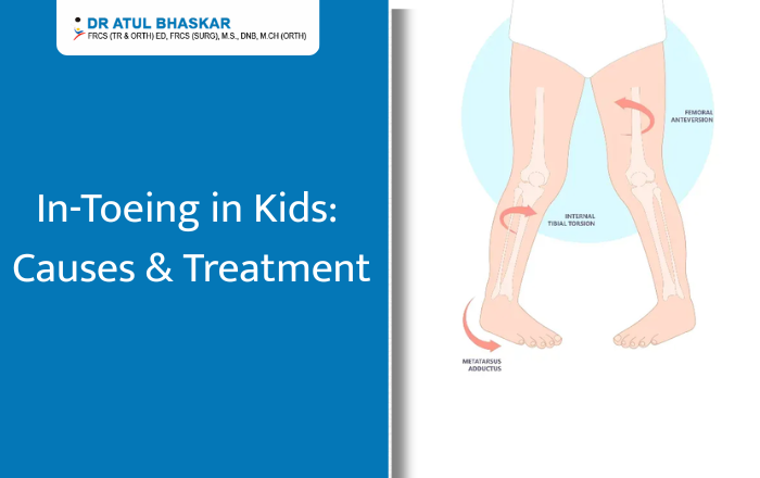
Pigeon Toes in Children: What Causes In-Toeing and How to Treat It
Pigeon Toes in Children: What Causes In-Toeing and How to Treat It Have you noticed your child walking or running with their feet pointing inward? This is known as pigeon toes or in-toeing, and it’s a common concern among parents. While it can look unusual, in many cases it’s simply a part of your child’s growth pattern and improves with time. However, sometimes it can be a sign that your child needs help to correct their walking style. In this blog, we’ll explain what pigeon toes are, what causes them, when to be concerned, and the different treatment options available. We’ll also share some home tips that may help your child’s feet grow in the right direction. What Are Pigeon Toes (In-Toeing)? Pigeon toes describe a walking pattern where a child’s feet point inward instead of straight ahead. This can happen in one foot or both. Parents usually notice it when their child starts walking, but it can also appear later in early school years. While it often corrects naturally, persistent or severe in-toeing should be checked by a doctor. Common Causes of In-Toeing in Children 1. Metatarsus AdductusThis condition appears in babies when the front part of the foot curves inward. It can happen because of the baby’s position in the womb. Mild cases often improve on their own, but stretching exercises may be suggested for some children. 2. Tibial TorsionIn toddlers, in-toeing can happen when the shinbone (tibia) naturally twists inward. This usually becomes noticeable when the child begins walking. As the child grows, the tibia often straightens out without treatment. 3. Femoral AnteversionThis occurs when the thigh bone (femur) twists inward, making the knees and feet turn in. It’s often most noticeable between ages 3 and 8. Many children grow out of it, but in some cases, it continues into later childhood. 4. Genetic and Developmental FactorsSome children have a family history of in-toeing. Growth patterns, sitting posture, and early walking habits can also influence how the feet and legs align. Symptoms and When to Seek Medical Advice Mild in-toeing is usually harmless. However, you should see a doctor if: Your child often trips or falls. The condition worsens over time. There is pain, limping, or swelling. One leg or foot looks very different from the other. Diagnosis: How Doctors Evaluate In-Toeing A pediatric orthopedic doctor will: Ask about your child’s growth and walking habits. Check leg and foot alignment during a physical exam. Observe your child’s gait while walking or running. Use X-rays or scans if needed to understand bone position. Treatment Options for Pigeon Toes 1. Observation and Natural CorrectionMost cases improve naturally as the bones and muscles develop. Doctors often recommend regular monitoring to track changes. 2. Stretching and ExercisesSpecific exercises can help improve muscle strength and foot alignment. These may include balance games, stretching routines, and walking on different surfaces. 3. Orthotics and Supportive FootwearIn some cases, special shoe inserts or braces are suggested to guide foot position. These are usually for children with more persistent symptoms. 4. Medical or Surgical InterventionSurgery is rarely needed, but in severe cases where in-toeing affects mobility or daily activities, it may be considered. Home Care and Prevention Tips Encourage barefoot walking indoors on safe, flat surfaces. Avoid letting your child sit in the “W” position. Include outdoor play and activities that strengthen leg and foot muscles. Make walking and balance games part of daily fun. Possible Complications if Left Untreated While many children experience no issues, untreated severe in-toeing can lead to: Frequent tripping or falling. Difficulty in sports or running. Increased strain on knees and hips over time. When to See a Pediatric Orthopedic Specialist You should consider a specialist if: Your child’s in-toeing persists beyond age 8. Pain or discomfort is present. The condition is affecting confidence or physical activities. Help Your Child Walk Confidently – Book a Consultation Today Most cases of pigeon toes in children are simply a stage of growth. Still, it’s always best to keep an eye on your child’s walking style and get professional advice if you notice ongoing problems. If you are concerned about your child’s in-toeing or walking pattern, don’t wait for it to worsen.For proper evaluation and treatment, you can consult Dr. Atul Bhaskar – Pediatric Orthopedic Surgeon in Mumbai. FAQ Q1: Is pigeon toe normal in toddlers?Yes, many toddlers have in-toeing, and it often improves naturally with age. Q2: Can pigeon toes cause problems later in life?Mild cases usually cause no issues, but severe cases may lead to tripping or joint strain. Q3: How can I help correct my child’s in-toeing at home?Encourage barefoot walking on safe surfaces, avoid W-sitting, and include balance activities in playtime.